
Fig. 1.
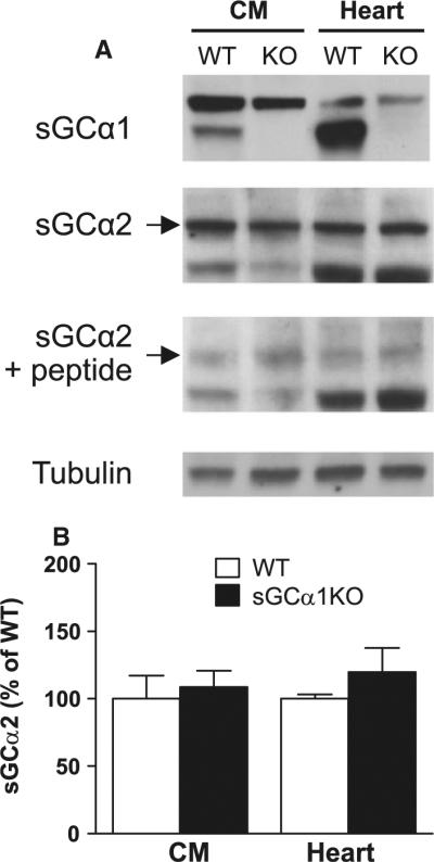
Immunoblot analysis of the sGCα1 and sGCα2 isoforms, as well as tubulin in whole hearts and cardiomyocytes of WT and sGCα1KO mice. a Representative immunoblots of sGC isoforms in homogenates of isolated cardiomyocytes (CM) and perfused whole hearts (heart). The sGCα2-immunoreactive band was blocked by the corresponding immunization peptide, confirming the specificity of the immunostaining. b Quantitative analysis of sGCα2 expression levels in WT (N = 4) and sGCα1KO (N = 5) samples, normalized to the respective WT samples. No significant differences were found between groups