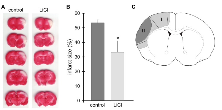
Fig. 1.
Administration of lithium reduced infarct size. A. Representative cerebral infarcts stained by a 2% TTC solution. Ischemic brains were harvested 48 h after stroke, sectioned into 5 blocks, and stained with TTC solution. Control, the group with control ischemia; LiCl, the group receiving both ischemia and lithium treatment. B. Infarct size of the ischemic cortex was measured, normalized to the contralateral cortex and expressed as a percentage according to the following formula: [(area of non-ischemic cortex - area of remaining ischemic cortex)/area of non-ischemic cortex] x 100. A mean value from the 5 blocks is presented. * P<0.05, vs. control. N=7/each group. C. Definition of ischemic core and penumbra. Tissue corresponding to the ischemic penumbra and core was dissected for the Akt kinase and Western blot assays. The gray region (I), including the black region (II), represents ischemic injury in a control rat with ischemia alone; the black region (II) represents infarction in a rat receiving ischemia plus lithium. The region spared by lithium is defined as the penumbra (region I) and the black area is defined as the ischemic core (region II). These regions were dissected for Western blot analysis. The corresponding non-ischemic cortex from a sham animal without ischemia was dissected for comparison.

