
Figure 4.
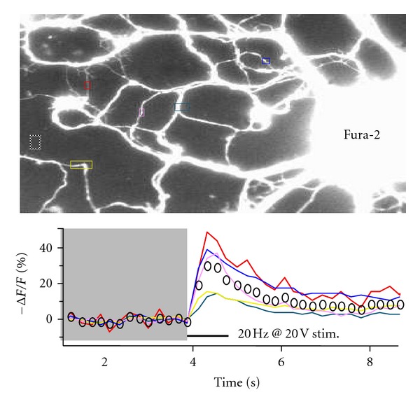
To confirm the viability of neurons transfected with BDNF-pH and their responsiveness to extracellular field stimulation, Ca2+ imaging was performed in neurons labeled with fura-2 AM. Upon electrical stimulation through field Pt wires, Ca2+ transiently increased in several dendrites. The graph shows the time course of background-subtracted delta F/F0 of 380 nm-excited fura-2 fluorescence intensity (510 nm emission) from the colored ROIs shown in the image. Images taken at 4 fps.