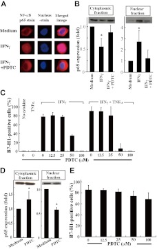
Figure 2.
Inhibition of NF-κB activation down-regulated B7-H1 expression.(A) SKM-1 cells cultured for 24 hours with medium alone, IFNγ, or IFNγ and pyrrolidine dithiocarbamate (PDTC; 100μM) were stained for NF-κB p65 (red) and nuclei (blue). The merged image showed that IFNγ induced p65 localization into the nucleus, which was inhibited by PDTC. Images were acquired on an Olympus IX71 inverted microscope using a LCPlanFI 40×/0.60 objective lens. Alexa 594 and 4'-6-diamidino-2-phenylindole were detected using Fluorescence Miler Units of U-MWIG2 and U-MNUA2 (Olympus), respectively. Images were captured using a 3CCD digital color camera (Hamamatsu Photonics) through Aquacosmos Software. (B) After the cultures in A, SKM-1 cells were separated into cytoplasmic and nuclear fractions, and their p65 contents were analyzed by Western blotting to show representative (top panel) and quantified (bottom panel) data (mean + SD) of 3 experiments. In each experiment, the band intensity of cell fractions from SKM-1 cells cultured with medium alone was defined as 1. *P = .017 and .06 (left panel) and *P = .045 and .012 (right panel) compared with “the medium” and IFNγ and PDTC, respectively. (C) SKM-1 cells cultured with or without cytokine(s) and various concentrations of PDTC were analyzed for B7-H1 expression by FCM. (D-E) Using F-36P cells expressing B7-H1 constitutively, the same experiments as in panels B and C were performed, except that no cytokines were used. Data are from 3 experiments. *P = .015 for D (left panel) and *P < .0001 (right panel) compared with the medium.

