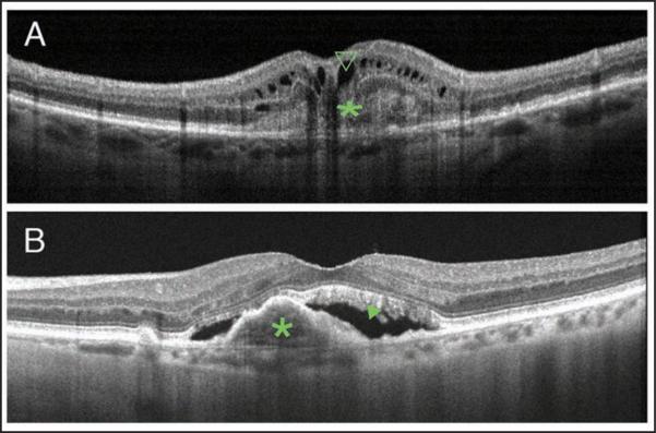
Figure 2.
Cross-sectional optical coherence tomography (OCT) images showing different types of choroidal neovascularization (CNV). (A) Cross-sectional image captured by RTVue (Optovue, Inc., Fremont, CA) showing a classic CNV, type 2 neovascularization, delineated as a nonuniform moderately hyperreflective formation above the RPE (green *) and the presence of intraretinal cysts (green open arrowhead). (B) Cross-sectional image captured by Heidelberg Spectralis HRA+OCT (Heidelberg Engineering, Heidelberg, Germany) showing a fibrovascular pigment epithelial detachment, type 1 neovascularization (green *), and the presence of subretinal fluid (green closed arrowhead).

