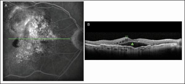
Figure 3.
Occult choroidal neovascularization membrane. (A) The fluorescein angiogram performed in Heidelberg Spectralis HRA+OCT (Heidelberg Engineering, Heidelberg, Germany) showed a speckled hyperfluorescence with dye pooled in the subretinal space on the late phase of the examination. (B) Cross-sectional image of Spectralis device depicted the thickened retinal pigment epithelium raised by non-uniform moderate hyperreflective formation with the presence of subretinal fluid (green closed arrowhead) and intraretinal fluid (green open arrowhead).

