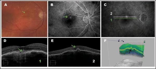
Figure 4.
Type 3 neovascularization (retinal angiomatous proliferation). (A) Color photography showing intraretinal hemorrhage (green arrow). (B and C) Fluorescein angiograms show poorly defined hyperfluorescence typical of retinal angiomatous proliferation (green arrow). (D) An enlarged cross-sectional Cirrus HD-OCT (Carl Zeiss Meditec, Inc., Dublin, CA) image showing a pigment epithelium detachment and a vessel penetrating the retina (green arrow). (E) Cross-sectional Cirrus HD-OCT image showing a pigment epithelial detachment, a hyperreflective material (green arrow) that can be the neovascular tissue, in the outer retina layers. (F) Three-dimensional Cirrus HD-OCT image showing a hyperreflective material in the outer retina layers (black arrow).

