
Figure 5.
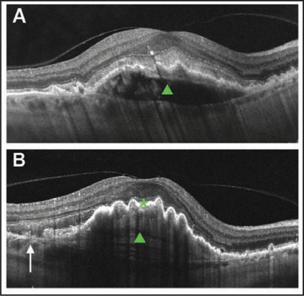
Cross-sectional Cirrus HD-OCT (Carl Zeiss Meditec, Inc., Dublin, CA) image of a fibrovascular pigment epithelium detachment (PED) before and after retinal pigment epithelium (RPE) rip. (A) Cross-sectional image before rip. Note dome-shaped PED (green closed arrowhead) and intact RPE layer. (B) Cross-sectional image after rip. Arrow denotes discontinuity of the RPE layer. Note RPE scrolling at the edge of the RPE defect and pleating RPE over the PED (green closed arrowhead).