
Figure 7.
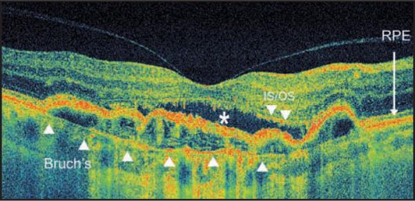
High pixel density high-speed ultra-high-resolution optical coherence tomography image from occult choroidal neovascularization. Note discontinuity of the inner/outer segment (IS/OS) junction line, subretinal fluid (white *), and pigment epithelium detachment. Bruch's membrane is demonstrated with the white arrowheads. RPE = retinal pigment epithelium.