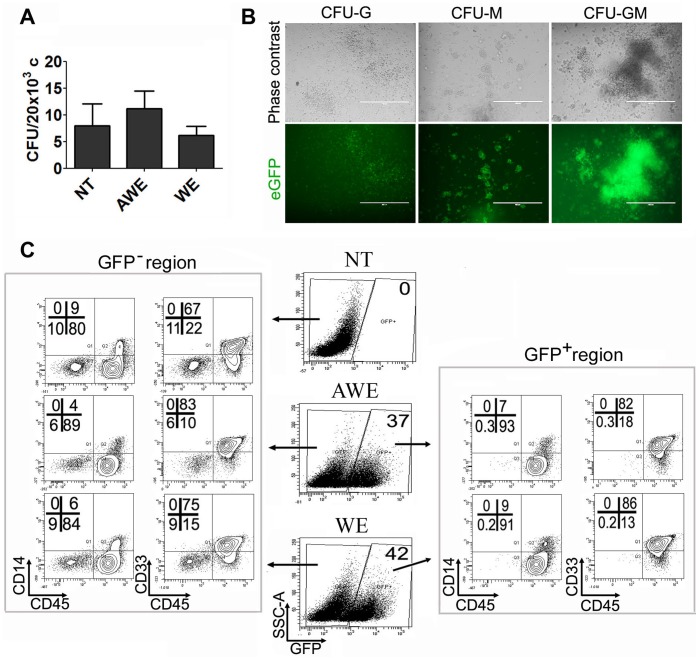
Figure 3. WAS-promoter driven LVs efficiently express eGFP in hESC-derived myeloid colonies.
AWE and WE-transduced hESCs were plated in EB hematopoietic differentiation media for 15 days and then seeded in methylcellulose H4434 (Stem Cell Technologies, Vancouver, Canada). (A) Graph showing CFU efficiency obtained from AWE and WE-transduced hESCs compared to untransduced hESCs (NT). Data are shown as average from three independent experiments +/− SEM. (B) Phase contrast (Top panels) and fluorescence (bottom panels) microphotographs from AWE-transduced hESCs derived CFUs. (C) Phenotypic analysis of cells derived from AWE and WE-transduced hESCs. eGFP+ (right plots) and eGFP− (left plots) populations were analyzed for expression of mature hematopoietic markers CD45, CD33 and CD14.

