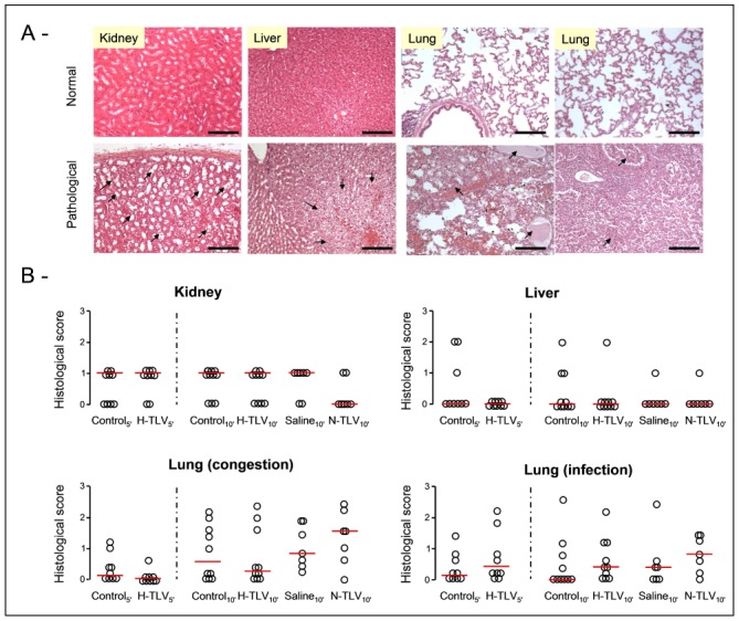
Figure 2.

Panel A: Examples of normal or pathological histological appearances of the kidney, liver and lungs in the TLV and control groups, respectively. In kidney, lesions consisted in dilated regenerative proximal tubules (arrows, bar=120 μm). In liver, we observed systematized clarification of hepatocytes (arrows, bar=120 μm). In lungs, lesions were congestion and serous edema (arrows in the left lung panel, bar=120 μm) or foci of bronchopneumonia (arrows in the right lung panel, bar=120 μm).
Panel B: Histological scores of alteration in kidney, liver and lungs of rabbits from the different groups. For lungs, we assessed two separate scores for cardiogenic lesions and infection complications, respectively. Open circles represents individual scores and the thick line represents the median value of corresponding group.
* p<0.05 vs corresponding control; TLV, total liquid ventilation; H-TLV, hypothermic TLV; N-TLV, normothermic TLV; Saline, hypothermia induced by intravenous administration of cold saline combined to external cooling.
