Figure 3.

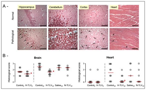
Panel A: Examples of normal or pathological histological appearances of the brain and the heart in the TLV and control groups, respectively. In brain, ischemic disorders consisted in ischemic pyramidal cells with pycnotic nucleus in the hippocampus (arrows, bar=30 μm), in laminar necrosis of Purkinje cells in the cerebellum (arrows, bar=30 μm) or in numerous ischemic neurons in the cortex (arrows, bar=30 μm), respectively. In the myocardium, we observed foci of cardiomyocytes necrosis (arrows, bar=120 μm).
Panel B: Histological scores of alteration in the brain and heart of rabbits from the different groups. Open circles represents individual scores and the thick line represents the median value of the corresponding group.
* p<0.05 vs corresponding control; TLV, total liquid ventilation; H-TLV, hypothermic TLV; N-TLV, normothermic TLV; Saline, hypothermia induced by intravenous administration of cold saline combined to external cooling.
