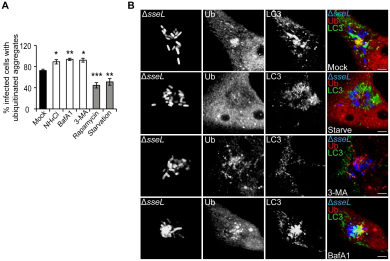
Figure 4. SCV-associated ubiquitinated aggregates are autophagy substrates.
(A) Quantification of the percentage of infected cells with SCV-associated ubiquitinated aggregates. HeLa cells were infected with ΔsseL mutant bacteria for 12 h. At 8 h post-infection, cells were subjected to the indicated treatments for 4 h before fixation and immunolabelling. A minimum of 50 cells were counted per condition in each experiment and values are the mean ± SEM of at least 3 independent experiments. p values are relative to the mean value of mock-treated cells. * p<0.05; ** p<0.01; *** p<0.001. (B) Single confocal sections of HeLa cells infected with GFP-expressing ΔsseL mutant bacteria (blue), treated and processed as in (A) and analysed by fluorescence microscopy for ubiquitin (Ub, red) and LC3 (green) (scale bars, 5 µm). The far right panels show merged images of LC3, ubiquitin and Salmonella.

