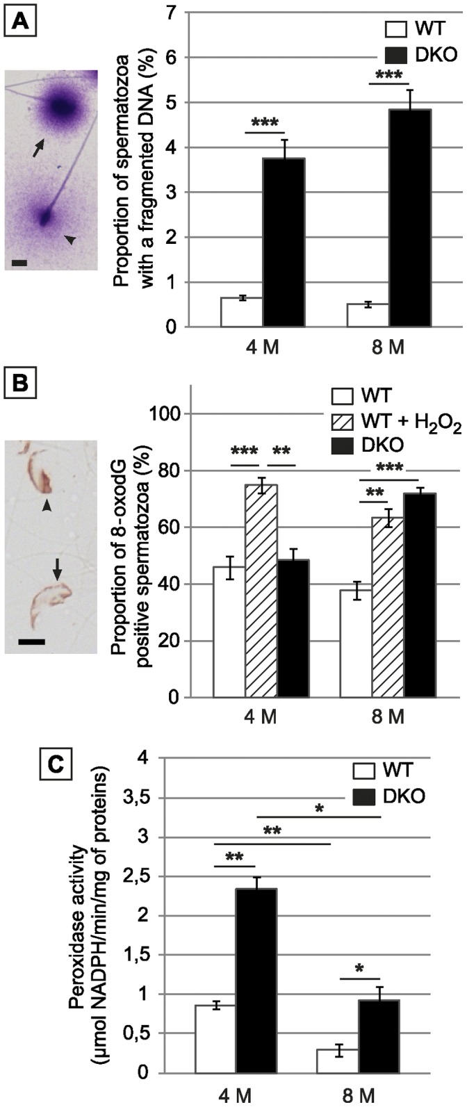
Figure 7. Cauda-retrieved spermatozoa of DKO animals suffer oxidative damage.

A: Left panel: Typical picture of fragmented (arrowhead) or non-fragmented (arrow) sperm nucleus as shown by the modified Sperm Chromatin Dispersion Assay. Scale bar = 5 µm. Right panel: Histograms show the proportion of WT and DKO cauda collected spermatozoa with a fragmented DNA in animals aged 4 or 8 months. B: Left panel: Typical immunodetection of the nuclear adduct 8-oxodG in cauda epididymidis-retrieved spermatozoa preparations from DKO male mice aged 8 months. Scale bar = 5 µm. Right panel: Histograms show the percentage of 8-oxodG positive spermatozoa in cauda epididymidis-retrieved spermatozoa preparations, respectively from WT, positive control (WT spermatozoa treated with H2O2) and DKO male mice aged 4 and 8 months. (Mean+/− SEM; n = 5; *: p≤0.05; **: p≤0.01; ***: p≤0.001). C: Histograms show global H2O2-scavenging activity using H2O2 as a substrate in cauda epididymidis tissue extracts from WT or DKO animals aged 4 or 8 months. (Mean +/− SEM; n = 5; *: p≤0.05; **: p≤0.01).
