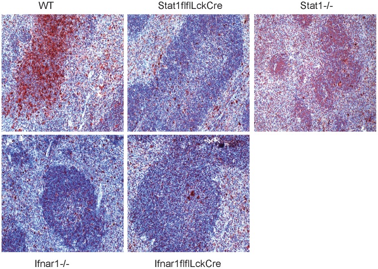
Figure 3. Apoptotic cell death in the spleen.
WT, LckCreStat1flfl, LckCreIfnar1flfl, Ifnar1−/− and Stat1−/− were infected with 1×10∧6 Lm and spleens were isolated 48 h after infection. TUNEL positive cells are visible in dark red; hematoxyline counterstaining indicates the structure of the spleen (3).

