
Figure 3. Compensatory YfiR alleles.
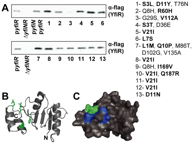
A) Immunoblots with M2 antiserum, showing levels of compensatory YfiR-flag variants in whole cell lysates. pyfiR: ΔyfiNR pMR-yfiR-flag, ΔyfiNR: strain without vector, pyfiN/pyfiR: ΔyfiNR pGm-yfiprom-N, pMR-yfiR-flag, lanes 1–13: ΔyfiNR pGm-yfiprom-N with compensatory pMR-yfiR-flag plasmids, proposed activating mutations are highlighted in bold. Mutants in lane 1, 4–8, 10–13 harbor mutations in the signal peptide that enhance expression, see also Table 2. B) Cartoon showing the locations of activating substitutions (green) on a homology model of YfiR (comprising residues 68–190). N and C termini are marked. The YfiR model is based on multiple structures (see Materials and methods). C) Surface representation of the YfiR model. The locations of activating mutants are shown in green, hydrophobic residues forming the possible YfiN binding surface are shown in dark blue.