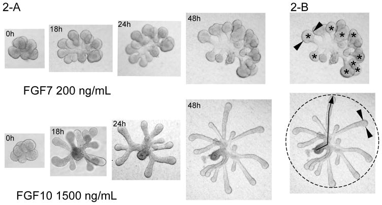
Figure 2.
SMG epithelial rudiments cultured with FGF7 or FGF10. Figure 2-A shows epithelial rudiments cultured for 48 h and the corresponding bud expansion or duct elongation promoted by FGF7 or FGF10, respectively. Figure 2-B illustrates how to quantitate morphological events during SMG epithelial morphogenesis. The upper figure shows how the width is measured (indicated by arrowheads) and the number (*) of end buds is counted; the bottom figure shows that the diameter (dotted circle) and the length of the ducts (arrow) can be measured.

