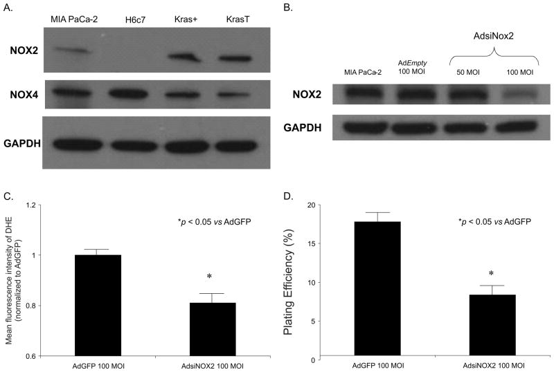
Figure 3. NOX2 is involved in superoxide production in pancreatic cancer cells.
A. Western blot analysis for NOX2 demonstrates expression in MIA PaCa-2, Kras+ and KrasT cells but no expression in the H6c7 pancreatic ductal epithelial cell line. NOX4 was present in all cell lines tested. GAPDH was used as a loading control
B. An adenoviral vector expressing siRNA against NOX2 (AdsiNOX2) was transfected into the MIA PaCa-2 pancreatic cancer cell line. At 100 MOI, there was a significant decrease in immunoreactive protein when compared to both the parental cell line and cells transfected the adenoviral control vector AdGFP (100 MOI).
C. MIA PaCa-2 cells infected with the AdsiNOX2 vector demonstrate a significant decrease in hydroethidine fluorescence when compared to the same cell line infected with the AdGFP vector. Mean fluorescence intensity was measured via flow cytometry, corrected for background fluorescence levels, and normalized to the cells infected with AdGFP. *p < 0.05 vs AdGFP cells, means ± SEM, n = 3.
D. AdsiNOX2 infection (100 MOI) in MIA PaCa-2 cells decreased plating efficiency when compared to the same cell line infected with the AdGFP vector (100 MOI). Each point represents the mean values, with p < 0.05 vs. AdGFP, n = 3.

