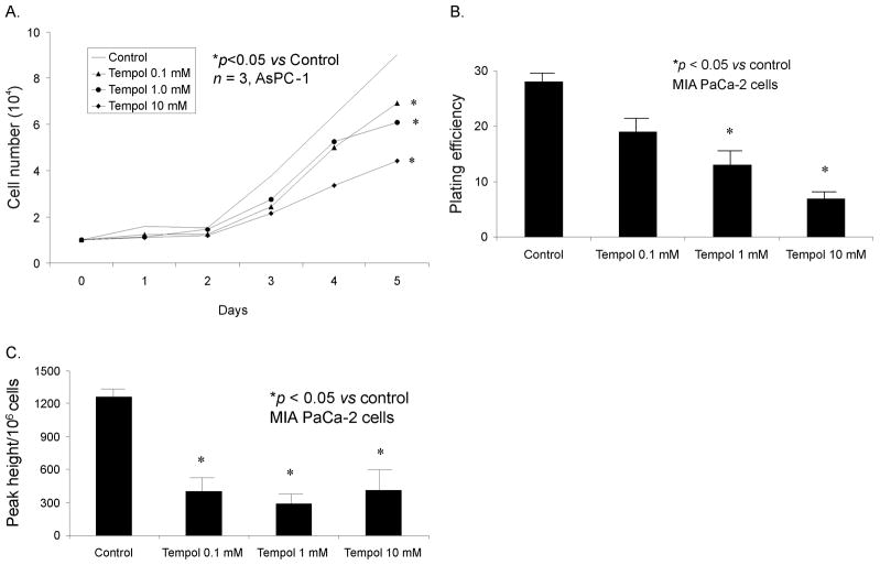
Figure 4. Tempol inhibits pancreatic cancer cell growth.
A. For cell growth, cells were treated with Tempol (0, 0.1, 1.0, and 10 mM) for 1 h. Mean in vitro cell growth of AsPC-1 cells are shown.
B. For clonogenic survival, cells were treated with Tempol (0, 0.1, 1 and 10 mM) for 1 h, 400 cells were plated in each well of 6-well plates and incubated at 37°C for 14 days. Colonies were fixed and stained with Coomassie blue; only colonies with more than 50 cells were counted. Each point was determined in triplicate from the same culture. * p < 0.01 vs control.
C. Electron paramagnetic resonance was performed in MIA PaCa-2 cells treated with Tempol to determine superoxide levels. EPR spectra were acquired and peak heights were quantified and compared in cells treated with Tempol (0.1–10 mM). DMPO-OH signals from MIA PaCa-2 cells treated with Tempol. In the spectral analysis the Tempol signals were removed to see the DMPO-OH signal better.

