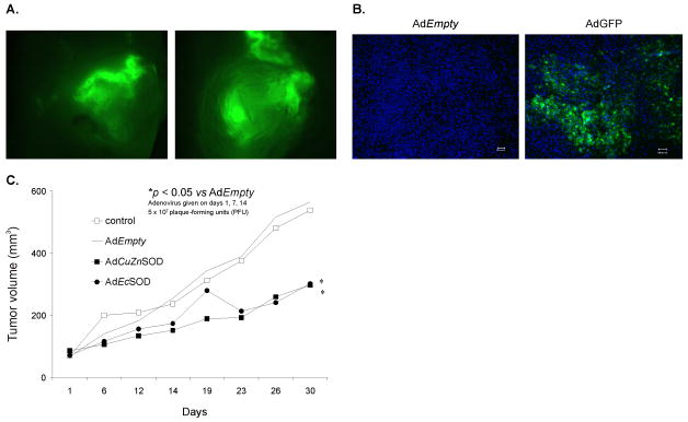
Figure 6. Intratumoral injections of AdCuZnSOD and AdEcSOD inhibit growth.
A. Detection of transgene expression and biodistribution in whole tumor xenografts. MIA PaCa-2 cells (2 × 10⁶) were injected subcutaneously into the flank region of nude mice and allowed the tumors to reach 4–5 mm in diameter. Tumors were then injected with AdGFP or AdEmpty adenoviral vectors. Two representative tumors demonstrate GFP expression which is clearly visible by the green fluorescence and heterogeneously distributed throughout the tumor.
B. Histological sections detecting transgene expression and biodistribution in MIA PaCa-2 human pancreatic tumor xenograft in vivo after intratumoral injection of an AdGFP construct (1 × 10⁹ PFU). AdEmpty was used as the control. Low power fields of fluorescence microscopy on sections from tumors injected with AdEmpty and AdGFP respectively. Sections were counterstained with DAPI. GFP expressing cells are clearly visible by their green fluorescence and are widely distributed throughout the tumor. Bar = 100 microns.
C. AdCuZnSOD or AdEcSOD injections decreased MIA PaCa-2 tumor growth in nude mice. The AdCuZnSOD and AdEcSOD groups had significantly slower tumor growth when compared to the AdEmpty group (p < 0.05, n = 6–8/group). MIA PaCa-2 tumor cells (2 × 10⁶) were delivered subcutaneously into the flank region of nude mice. Controls received serum-free media in similar volumes. 5 × 10⁷ PFUs of the AdCuZnSOD, AdEcSOD, or AdEmpty constructs were delivered to the tumor on days 1, 7 and 14 of the experiment. On day 30 there was nearly a 2-fold decrease in tumor growth in animals receiving the AdEcSOD vector or AdCuZnSOD when compared to treatment with the AdEmpty vector.

