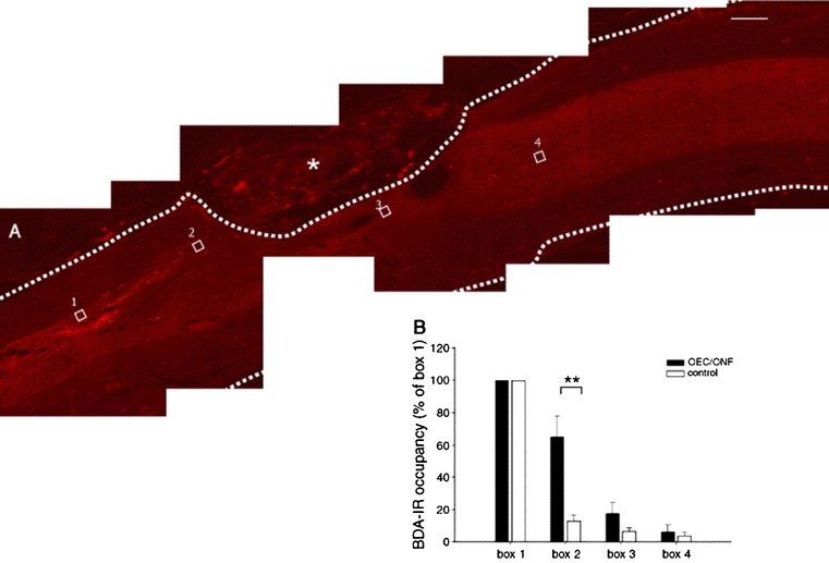
Fig. 4.
Biotinylated dextran amine (BDA)-labeled corticospinal tract axons and response to OEC/ONF intervention. BDA-labeled corticospinal axons were observed to approach the graft/lesion site, but only very few grew underneath the lesion and into the caudal host spinal cord. No labeled corticospinal axons penetrated the graft/lesion site. a Sagittal spinal cord section of an OEC/ONF-transplanted animal. The dotted line represents the delineation of the spinal cord; the OEC/ONF–biomatrix complex is visible within the lesion site. The four boxes represent the boxes used in the quantitative analysis. b A significantly higher BDA-labeling is present directly rostral to the injury site (box 2) in transplanted animals versus control animals. Scale bar 200 um. Adapted from Deumens et al. (2006), with permission

