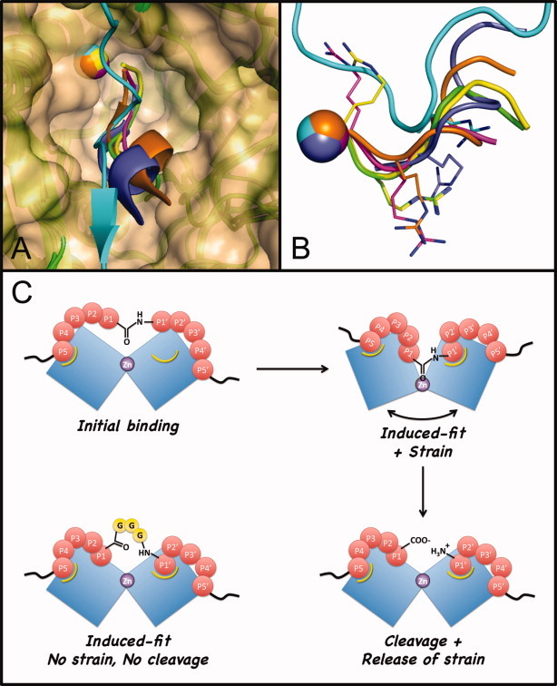
Figure 3.

Active-site plasticity of BoNT/A-LC. A: Shown are superposed structures of BoNT/A-LC(E224Q/Y366F) mutant complexed with SNAP25(141-204) (pdb1xtg, cyan) and 5 wildtype BoNT/A-LC complexes with Ac-CRATKML (pdb3boo, purple); I1 peptide (pdb3ds9, orange); QRATKM-NH2 (pdb3dda, green); RRATKM-NH2 (pdb3ddb, yellow); or RRGC-NH2 (pdb3c88, magenta). Zinc ions are shown as spheres. The surface at the BoNT/A-LC active site was generated from the pdb1xtg structure. B: Shown are the main chains of the pseudosubstrates or peptidomimetics from the same overlaying structures as in (A) after a 90° rotation. Side chains of all Arg residues are shown as sticks. The graphic image was generated with Pymol using the corresponding Protein Data Base (PDB) files. C: An induced-fit model for BoNT/A-LC-catalyzed cleavage of SNAP25 and inhibition of BoNT/A-LC by the Gly-insertion SNAPI (3G). Blue blocks represent enzyme surface at the catalytic cleft. Pockets marked with yellow curves are the induced, high affinity sites for P5 Asp and P1′ Arg.
