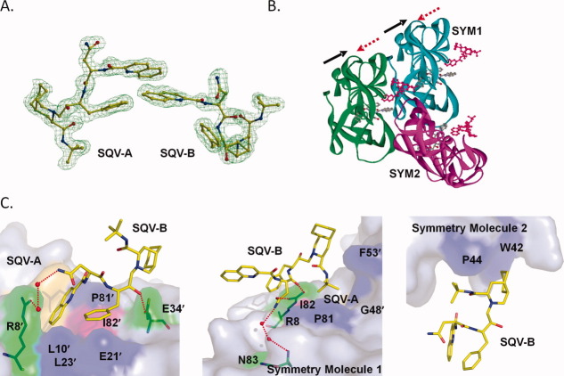
Figure 3.
SQV-B in the extended binding site of PR1M. A: Omit map for SQV-A and SQV-B. SQV-B interacts with SQV-A in an extension of the regular active site cavity. B: SQV-B molecule is surrounded by three PR1M dimers in the crystal lattice, colored in green, cyan, and magenta. The SQV-A occupying the regular binding pocket is colored by atom type, whereas the extra SQV-B is shown in red. The arrows represent the alternating orientations of SQV-A and SQV-B molecules. C: SQV-B interactions with PR1M dimer, symmetry-related dimer 1, and symmetry-related dimer 2. Mutated residues are in pink surface representation, SQV-A is in golden color, and the surfaces of the residues involved in polar or hydrophobic interactions are shown in green and blue, respectively. Water molecules are shown as red spheres. Hydrogen bonds are indicated by red dotted lines. [Color figure can be viewed in the online issue, which is available at wileyonlinelibrary.com.]

