
Figure 5.
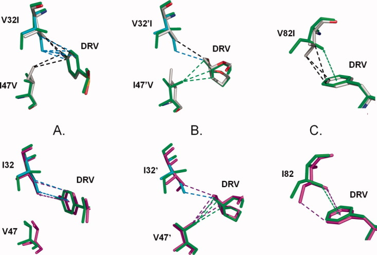
Structural comparison of DRV complexes with PR1M, PR1, and PR2. The PR1 structure is colored by atom type, PR1M is shown in green bonds with the minor conformations of Ile32 and 32′ in cyan, and PR2 is colored magenta. Only the major conformation of DRV is shown for PR1-DRV. Hydrophobic interactions are indicated as dashed lines. CH…π interactions are indicated by dashed arrows. PR1M is compared with PR1 in the upper panels and with PR2 in the lower panels. A: Residues 32 and 47 interactions with P2′ aniline of DRV; B: Residues 32′ and 47′ interactions with bis-THF at P2; and C: Residue 82 interactions with P1 of DRV. [Color figure can be viewed in the online issue, which is available at wileyonlinelibrary.com.]