
Figure 6.
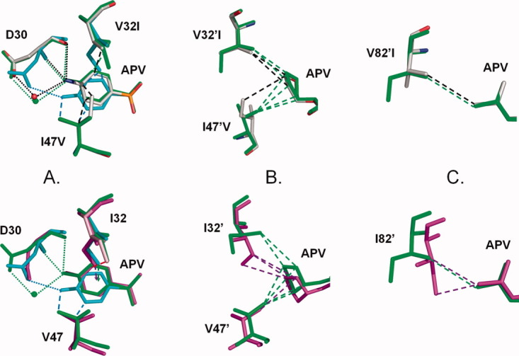
Comparison of PR1, PR2, and PR1M interactions with APV. The PR1 structure is colored by atom type, PR1M is shown in green bonds with the minor conformations of Ile32 and 32′ in cyan, and PR2 is colored magenta. Only the major (0.7 occupancy) conformation is shown for APV and PR1 residues in the wild type complex. Hydrogen bond interactions are shown as dotted lines, hydrophobic interactions are indicated as dashed lines, and CH…π interactions are indicated by dashed arrows. A: Asp30, residues 32 and 47 interactions with aniline of APV. The aniline group and Asp30 have two alternate conformations in PR1M -APV. The major conformations closely resemble the wild type structure with strong hydrogen bond interactions. The minor conformation (in cyan) has only one hydrogen bond and more hydrophobic interactions with Val47. B: Residue 32′ and 47′ interactions with the THF group of APV. C: Residue 82′ interactions with the P1′ group of APV. [Color figure can be viewed in the online issue, which is available at wileyonlinelibrary.com.]