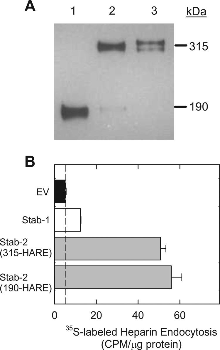
FIGURE 2.
Internalization of 3-O-sulfated heparin via Stab1 and Stab2. A, cell lysates (20 μg) were separated by 5% SDS-PAGE, blotted to nitrocellulose, and probed with anti-V5 antibody. Lane 1, Stab2/190-HARE; lane 2, Stab2/315-HARE; lane 3, Stab1. B, stable cell lines expressing Stab1 (white bar), both Stab2 isoforms (gray bars), and empty vector (EV, black bar) were incubated with 35S-labeled 3-O-sulfated heparin for 3 h. The dotted line represents nonspecific binding values and is subtracted from the data to determine receptor-specific endocytosis. Endocytosis was evaluated by cpm/μg of cell lysate protein for each cell line, mean ± S.D., n = 3.

