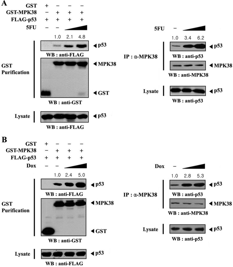
FIGURE 3.
Modulation of the association between MPK38 and p53 by p53 signals. HEK293 cells transfected with the indicated expression vectors were incubated with or without increasing amounts of 5-FU (0.38 and 0.57 mm, 30 h) or doxorubicin (Dox) (100 and 150 ng/ml, 24 h) as described previously (23). Cell lysates were purified on glutathione-Sepharose beads (GST purification), followed by immunoblot analysis using an anti-FLAG antibody to determine the complex formation between MPK38 and p53 (A and B, left panels). The endogenous level of MPK38-p53 complex in the presence or absence of 5FU (or doxorubicin) was also analyzed by immunoblotting with an anti-p53 antibody (A and B, right panels). The relative level of MPK38-p53 complex formation was quantitated by densitometric analysis, and the fold increase relative to untreated control cells expressing MPK38 and p53 (A and B, left panels) or untreated HEK293 cells (A and B, right panels) was calculated. IP, immunoprecipitation; WB, Western blot.

