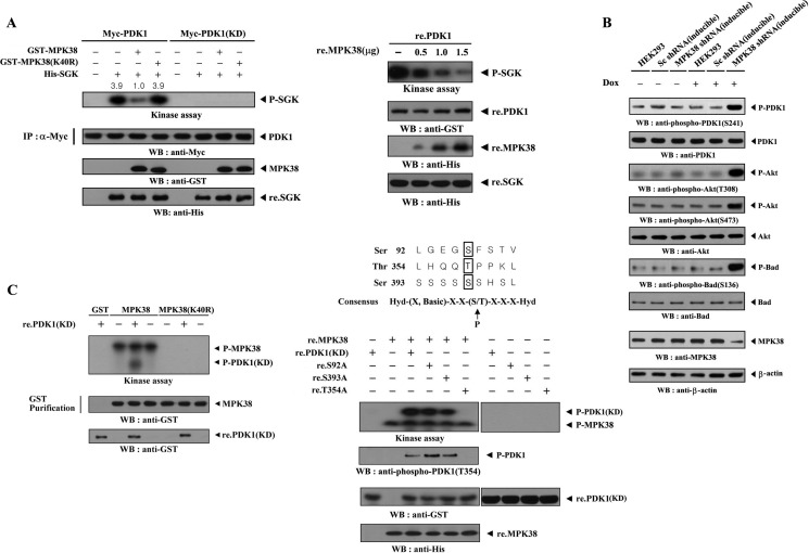
FIGURE 3.
MPK38 inhibits PDK1 kinase activity. A, HEK293 cells were transiently transfected with Myc-tagged wild-type or kinase-dead PDK1 in the presence or absence of GST-tagged wild-type and kinase-dead Mpk38. Cell lysates were subjected to immunoprecipitation (IP) with an anti-Myc antibody, and the Myc immunoprecipitates were analyzed for PDK1 kinase activity in an in vitro kinase assay using recombinant SGK as the substrate (left panel). Purified recombinant PDK1 proteins (re.PDK1) were incubated at room temperature for 1 h with the indicated amount of recombinant MPK38 (re.MPK38) in 50 μl of 50 mm HEPES buffer, pH 7.4, and then analyzed for PDK1 kinase activity in an in vitro kinase assay (right panel). P-SGK indicates phosphorylated SGK. B, effect of MPK38 on PDK1 downstream signaling. HEK293 cells harboring the stably integrated pSingle-tTS-shRNA vector containing scrambled shRNA (inducible Sc shRNA) or the pSingle-tTS-shRNA vector containing Mpk38-specific shRNA (inducible MPK38 shRNA), or parental HEK293 cells, were cultured in the presence or absence of 1 μg/ml doxycycline (Dox) for 72 h, and PDK1 downstream signaling was determined by immunoblot analysis. Inducible silencing of endogenous MPK38 by doxycycline was assessed by immunoblotting using an anti-MPK38 antibody. β-Actin was used as a loading control. C, identification of MPK38 phosphorylation sites on PDK1. After 48 h of transfection with GST-tagged wild-type or kinase-dead Mpk38, HEK293 cell lysates were subjected to precipitation with glutathione-Sepharose beads (GST purification) and analyzed in an in vitro kinase assay with recombinant kinase-dead PDK1 as the substrate (left panel). The in vitro kinase assay was performed with ∼3 μg of recombinant GST-tagged kinase-dead PDK1 or one of its substitution mutants (S92A, T354A, or S393A) in the presence or absence of recombinant His-tagged wild-type MPK38 (∼6 μg) (right panel). The purity of recombinant MPK38 and PDK1 proteins used for this experiment was confirmed by Coomassie Blue staining (see supplemental Fig. 4B). P-MPK38 and P-PDK1(KD) indicate autophosphorylated MPK38 and phosphorylated kinase-dead PDK1, respectively.

