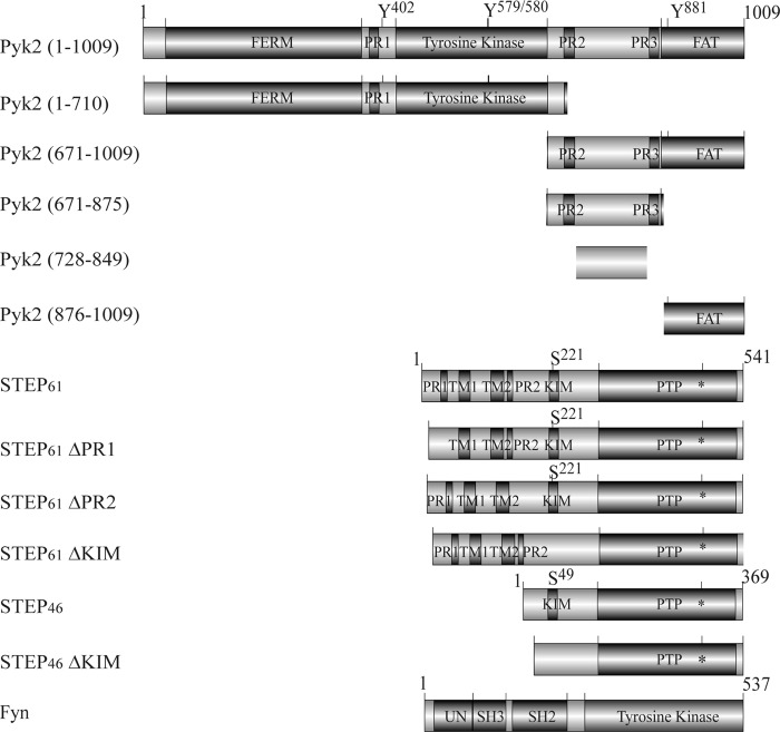
FIGURE 1.
Schematic domain structures used in this study. FERM, band 4.1/ezrin/radixin/moesin homology domain; FAT, focal adhesion-targeting domain; TM1 and TM2, putative transmembrane domains; PTP, protein-tyrosine phosphatase domain; asterisk, cysteine residue in the catalytic core; UN, unique N-terminal domain. Regulatory phosphorylation sites are shown. Numbering refers to amino acid residues within the proteins.

