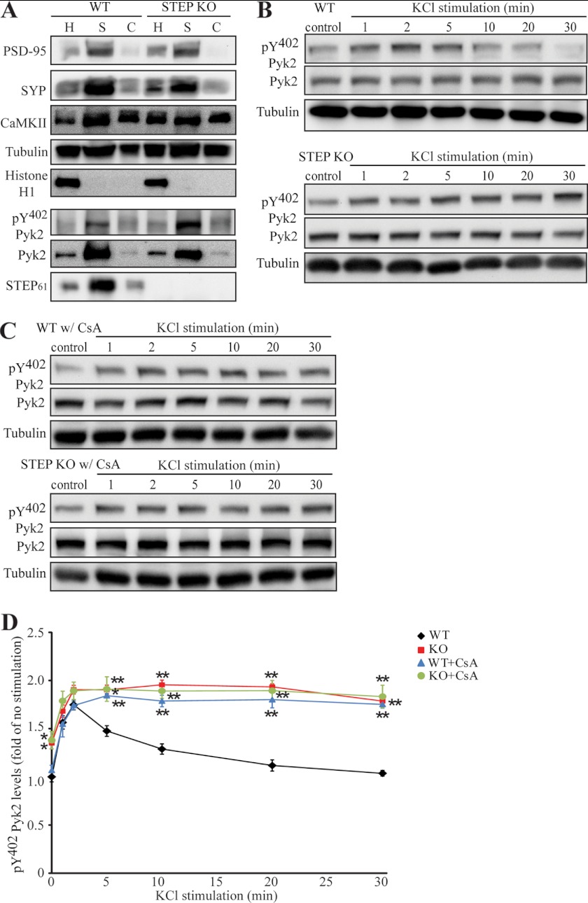
FIGURE 10.
Activation of STEP leads to dephosphorylation of Pyk2 in synaptoneurosomes. A, expression of presynaptic and postsynaptic markers in synaptoneurosome preparation. SYP, synaptophysin; CaMKII, Ca2+/calmodulin-dependent protein kinase II. B, synaptoneurosomes from WT (upper panel) or STEP KO (lower panel) mice were stimulated with 40 mm KCl for the indicated durations. Phosphorylation of Pyk2 was determined with Tyr(P)402 Pyk2 antibody. C, CsA-pretreated synaptoneurosomes (100 nm; 10 min) from WT (upper panel) or STEP KO (lower panel) mouse brains were stimulated with 40 mm KCl as in B. D, quantitation of phospho-Pyk2 from B and C. Phosphorylation levels were first normalized to total protein and then to tubulin as a loading control. Error bars indicate the SEM of at least three independent experiments. All values were expressed as -fold changes compared with WT control levels (*, p < 0.05; **, p < 0.01; one-way ANOVA with post hoc Tukey test; n = 4).

