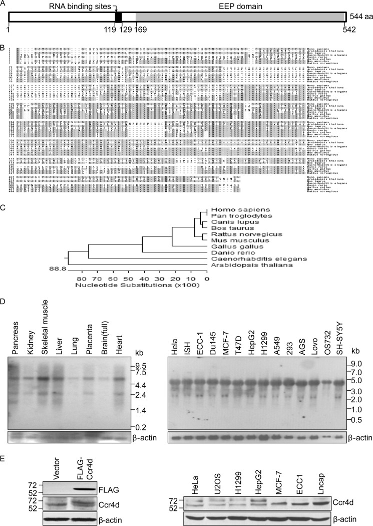
FIGURE 1.
Cloning, characterization, and expression of Ccr4d. A, a schematic representation of the structure of Ccr4d. A conserved domain (EEP domain) and putative RNA binding sites are shown. aa, amino acids. B, amino acid sequence alignment of Ccr4d from different species. Shaded residues represent conserved regions. C, phylogenetic analysis of evolutionary relationships among homologues of Ccr4d proteins from different species. D, Northern blotting analysis of ccr4d mRNA expression in normal human tissues and in various tumor cell lines. β-actin was used as a loading control. E, Western blotting analysis of Ccr4d protein expression. Left, MCF-7 cells were transfected with empty vector or FLAG-Ccr4d constructs. Cellular proteins were prepared, and Western blotting was performed with anti-FLAG or anti-Ccr4d antibody. Right, Ccr4d expression in a collection of tumor cell lines was analyzed using anti-Ccr4d antibody. β-actin was used as a loading control.

