
FIGURE 2.
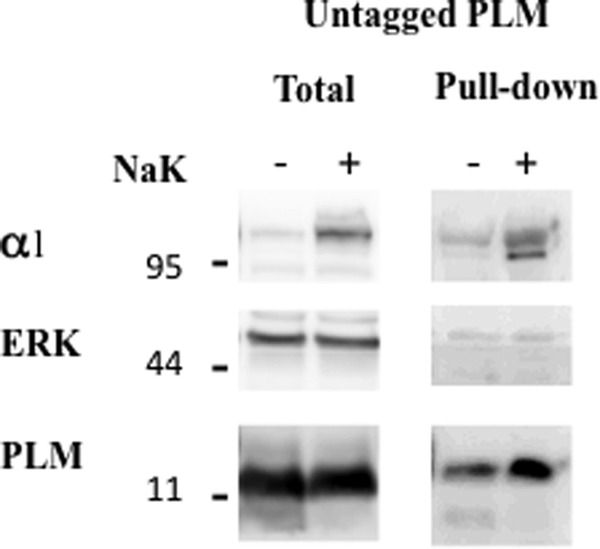
Cell surface biotinylation of PLM. Oocytes were injected with 2 ng of unmodified PLM ± 10 ng α1 and 7 ng β1 Na+/K+ ATPase (NaK). Three days later, oocytes were biotinylated by a 60-min incubation with 0.5 mg/ml DBB at room temperature. Oocytes were lysed, and biotinylated proteins were precipitated on streptavidin beads as described under “Experimental Procedures.” Total and pulled-down biotinylated proteins were resolved electrophoretically, and the blot was cut into low, medium, and high MW regions that were blotted for PLM, ERK, and α1 Na+/K+ ATPase.