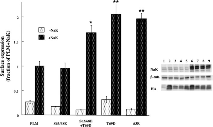
FIGURE 4.
Mutating Thr-69 and truncating C tail arginine residues elevate surface expression of PLM. Oocytes were injected with cRNA mixtures coding for the indicated PLM constructs (2 ng) ± α1 (10 ng) and β1 (7 ng) Na+/K+ ATPase (NaK). Left panel, surface expression of the HA epitope under the different conditions. The chemiluminescence signal was normalized to the total unmutated PLM protein expressed, and data are expressed as fractions of the mean value in oocytes injected with PLM+NaK (means ± S.E. of 16 oocytes). Probabilities for differences in surface expression between mutated and unmutated PLM were calculated using unpaired Student's t test. *, p < 0.0005; **, p < 0.0002. Right panel, Western blot analyses of the same oocytes with antibodies to HA, β tubulin, and α1Na+/K+ ATPase. The different lanes represent oocytes injected with the following cRNA mixtures: Lane 1, non-injected; lane 2, PLM; lane 3, S63/68E, lane 4, S63/68E+T69D; lane 5, Δ3R; lanes 6–9, as lanes 2–5 plus αβ Na+/K+ ATPase.

