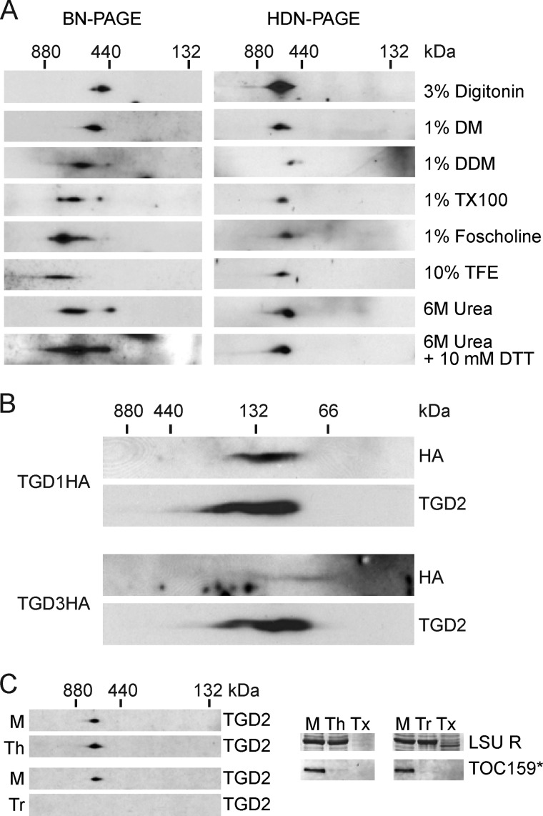
FIGURE 2.
The stable TGD1, -2, and -3 complex is present in the chloroplast inner envelope. A, anti-TGD2 immunoblots of two-dimensional gels in which the first dimension is a 4–10% BN-PAGE (left) or HDN-PAGE (right) and the second dimension is a 12% SDS-PAGE. Isolated wild-type Arabidopsis chloroplasts (equivalent to 20 μg of chlorophyll) were solubilized with reagents listed at the right (DM, decylmaltoside; TX100, Triton X-100; TFE, trifluoroethanol). In the case of trifluoroethanol and urea samples, 1% DDM was also present. B, immunoblots detecting proteins indicated at the right, similar to the HDN-PAGEs in A, except the gradient was 4–14%, and chloroplasts isolated from Arabidopsis of genotypes indicated at the left were solubilized in 1% SDS. C, anti-TGD2 immunoblots similar to the HDN-PAGE solubilized with DDM in A, except prior to solubilization the chloroplasts were mock-treated (M), thermolysin-treated (Th), or trypsin-treated (Tr). Immunoblots of single-dimension SDS-PAGEs show digestion of the 86-kDa fragment of TOC159 (TOC159*), and Coomassie Brilliant Blue stain shows protection of the large subunit of Rubisco (LSU R). Tx, trypsin treatment with Triton X-100 present.

