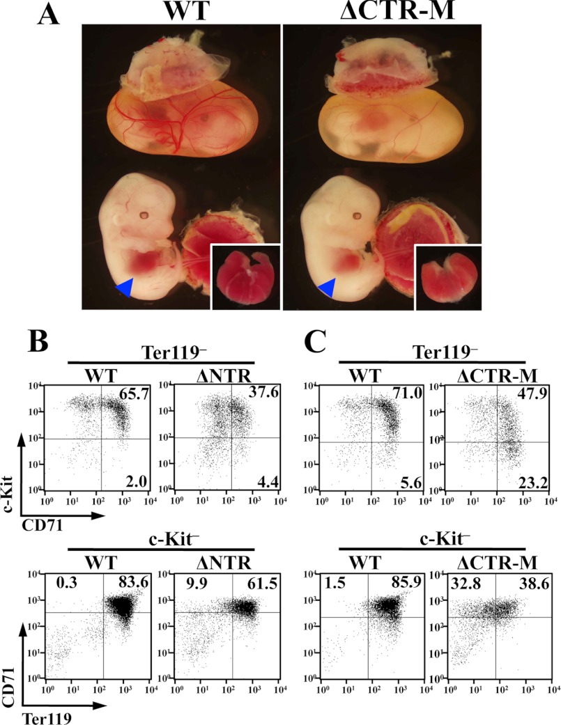
FIGURE 4.
Impaired erythroid differentiation in ΔCTR embryos. A, ΔCTR-M embryo (right panel) is distinguished from its wild-type (WT) littermate (left panel) by the anemic appearance. Small and pale livers of ΔCTR-M embryos are shown in the inset. B and C, flow cytometry evaluation of erythroid differentiation. Fetal liver monoclonal cells of E13.5 embryos of ΔNTR (B) and ΔCTR-M (C) were analyzed with the expressions of c-Kit and CD71 (upper panels) or CD71 and Ter119 (lower panels) in a Ter119-negative or c-Kit-negative fraction, respectively. Wild-type littermate was used as control.

