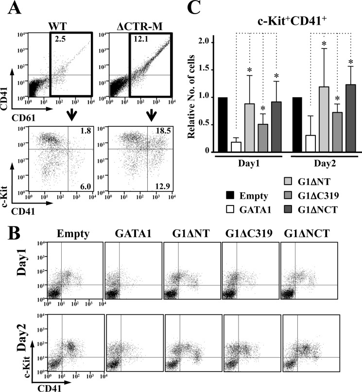
FIGURE 7.
Both N-TAD and C-TAD are involved in the GATA1-mediated megakaryocyte growth control. A, flow cytometry analysis of E13.5 fetal livers. CD41+CD61+ cells are abundant in ΔCTR-M embryo (upper panels). Note that c-Kit+CD41+CD61+ cells are dominantly increased in number. B and C, anti-proliferative activity of GATA1 for GATA1-deficient megakaryocytes is attenuated by C-TAD deletion, as is the case for N-TAD. Representative flow cytometry analysis (B) and statistical examination of the number of c-Kit+CD41+ cells from four independent experiments at day 1 and day 2 (C) are shown. The value in the cells introduced empty vector is set to one in every experiment in C. Data are presented as mean ± S.D. (*, p < 0.05).

