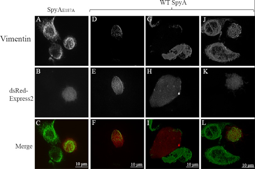
FIGURE 8.
Vimentin filamentation in SpyA-transfected RAW 264.7 cells. RAW cells were transfected with the mammalian expression vector pEF1α-IRES-DsRed-Express2-SpyAE187A, expressing dsRed-Express2 and either wild type SpyA or SpyAE187A bicistronically. 18 h after expression, the cells were stimulated with 100 ng/ml LPS to induce vimentin expression and filamentation, fixed, permeabilized, and stained for vimentin. Transfectants were identified by the expression of intracellular fluorescent protein dsRed-Express2. A–C, transfected cell expressing dsRedExpress/SpyAE187A stained for vimentin. Staining was identical to that of control (non-transfected, mock-transfected, or empty vector) cells. D–F, a subset of SpyA-transfected cells had only partial vimentin staining. G–I, the majority of wild type SpyA-transfected cells exhibited little to no vimentin staining, some of which exhibited a flattened morphology. J–L, a minority of cells transfected with the wild type SpyA expression vector pEF1α-IRES-DsRed-Express2-SpyA were consistent with and indistinguishable from control cells. 28 of 35 cells transfected with the wild type SpyA exhibited decreased or no vimentin staining compared with 4 of 39 cells transfected with the SpyAE187A catalytic mutant (p < 0.0001).

