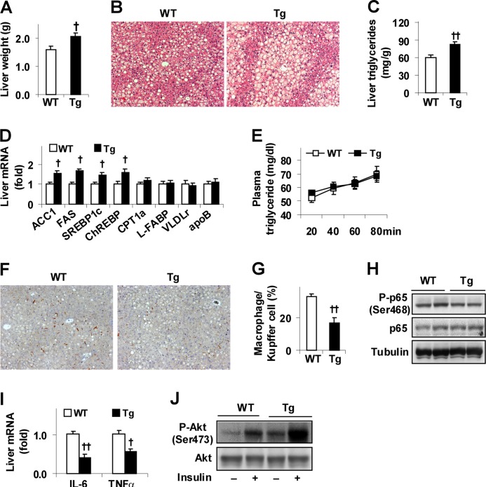
FIGURE 3.
Overexpression of PFKFB3/iPFK2 in adipose tissue decreases diet-induced liver inflammatory response and improves insulin signaling while increasing hepatic steatosis. At 5–6 weeks of age, male Tg and WT mice were fed an HFD for 12 weeks. A, changes in liver weight. B, liver histology. Liver sections were stained with hematoxylin and eosin (10×). C, levels of hepatic triglycerides were quantified using the biochemical assay. D, liver gene expression was quantified using real time RT-PCR. E, VLDL-triglyceride secretion. HFD-fed mice were fasted for 5 h and injected with tyloxapol (500 mg/kg, i.v.). Plasma levels of triglycerides were measured in blood samples taken at the indicated time points after the injection. F, liver macrophages/Kupffer cells. Liver sections were immunostained for F4/80. G, fraction of liver macrophages/Kupffer cells. H, liver inflammatory signaling. The levels of NF-κB p65 and phospho-p65 (Ser-468) were examined using Western blot analyses. I, liver expression of proinflammatory cytokines was quantified using real time RT-PCR. J, liver insulin signaling. Tissue samples of HFD-fed mice were collected at 5 min after a bolus injection of insulin (1 unit/kg) or PBS into the portal vein. The levels of Akt1/2 and phospho-Akt (Ser-473) were examined using Western blot analyses. A, C–E, G, and I, data are means ± S.E., n = 6–10. †, p < 0.05; ††, p < 0.01 Tg versus WT (A, C, and G) for the same gene (D and I).

