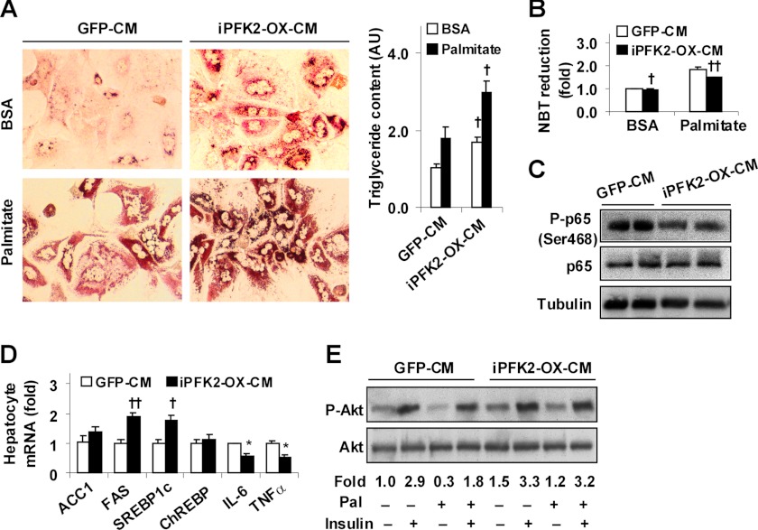
FIGURE 6.
Adipocyte factors generated in response PFKFB3/iPFK2 overexpression decrease hepatocyte inflammatory response and improve insulin signaling while increasing hepatocyte fat deposition. Wild-type mouse primary hepatocytes were treated with adipocyte-CM in M199 medium at a 1:1 ratio for 48 h in the presence or absence of supplemental palmitate (250 μm) for the last 24 h. Each of the assays was performed at least in quadruplicate. A, adipocyte-CM-treated mouse primary hepatocytes were stained with Oil Red O for 1 h (left four panels) and/or subjected to quantification of triglyceride content (right panel). AU, arbitrary unit. B, hepatocyte ROS production. C, hepatocyte levels of NF-κB p65 and phospho-p65 (Ser-468). D, hepatocyte gene expression. E, hepatocyte insulin signaling. A, B, and D, numeric data are means ± S.E. †, p < 0.05; ††, p < 0.01 iPFK2-OX-CM versus GFP-CM under the same condition (palmitate or BSA in A and B) and for the same gene (D). E, hepatocytes were incubated with or without insulin (100 nm) for 30 min prior to harvest. Phospho-Akt (Ser-473) to Akt1/2 ratios were calculated using densitometry and expressed as fold changes. Pal, palmitate.

