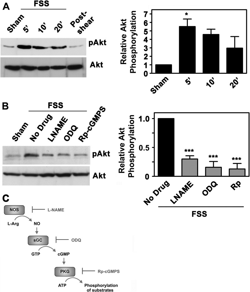
FIGURE 1.
FSS-induced Akt activation requires NO/cGMP/PKG signaling. A, serum-deprived hPOBs were mounted in a parallel plate flow chamber and kept under static conditions for 30 min (sham-treated; lane 1) or were subjected to laminar FSS at 12 dynes/cm2 for 5 min (lane 2), 10 min (lane 3), or 20 min (lanes 4 and 5); cells in lane 5 were kept in the flow chamber for an additional 10 min after cessation of flow. Western blots were analyzed with antibodies specific for Akt phosphorylated on Ser473 (pAkt; upper panel) or total Akt (lower panel) as described under “Experimental Procedures.” The bar graph on the right represents relative intensities of the pAkt bands normalized to total Akt, as measured by densitometry (mean ± S.E. (error bars) of three independent experiments; *, p < 0.05 for the comparison between sham- and shear-treated cells). B, hPOBs were preincubated for 1 h in the presence of 4 mm l-NAME, 10 μm ODQ, or 100 μm (Rp)-8-pCPT-PET-cGMPS, as indicated, and then exposed to FSS for 5 min or kept static (sham). Relative Akt phosphorylation was measured by Western blotting and densitometry as described in A, but the amount of pAkt in shear-stressed cells preincubated with medium only was assigned a value of 1 (***, p < 0.001 for the comparison between drug-treated cells and cells treated with medium only). C, the NO/cGMP/PKG signaling pathway with pharmacological inhibitors: l-NAME for NO synthase (NOS), ODQ for soluble guanylate cyclase (sGC), and (Rp)-8-pCPT-PET-cGMPS (Rp-cGMPS) for PKG.

