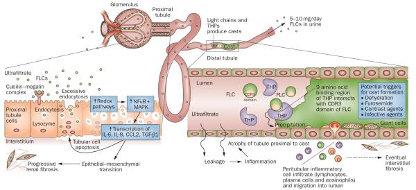
Figure 1.
Mechanisms of FLC-induced acute kidney injury. The very high concentrations of FLCs present in the ultrafiltrate of patients with multiple myeloma can result in direct injury to PTCs. The excessive endocytosis of FLCs by the cubilin–megalin complex expressed on PTCs can trigger apoptotic, proinflammatory and fibrotic pathways. Activation of redox pathways occurs, with increased expression of NFκB and MAPK, which in turn leads to the transcription of both inflammatory and profibrotic cytokines, such as IL-6, IL-8, CCL2 and TGF-β1. In the distal tubules, FLCs can bind to a specific binding domain on THPs and co-precipitate to form casts. These casts result in tubular atrophy proximal to the cast and lead to progressive interstitial inflammation and fibrosis. Abbreviations: CCL2, C-C motif chemokine 2; CDR, complementarity determining region; FLC, free light chain; IL, interleukin; MAPK, mitogen-activated protein kinase; NFκB, nuclear factor κB; PTC, proximal tubule cell; TGF-β1, transforming growth factor β1; THP, Tamm-Horsfall protein.

