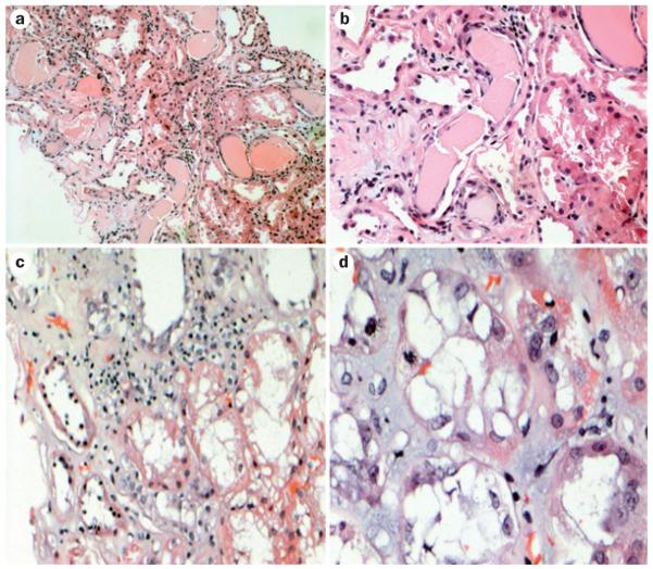
Figure 4.
Renal injuries induced by free light chains in patients with acute kidney injury. All samples were stained with hematoxylin and eosin. The classic appearance of cast nephropathy can be seen in a | and b | (magnification ×150 and ×350, respectively). Casts with fracture planes in distal nephrons and surrounding reactive tubular cells can be seen (center). Note adjacent proximal tubular damage and mild inflammation surrounding the casts. c | Marked tubular damage is observed, with inflammatory infiltrate that contains a mixture of mononuclear inflammatory cells and scattered eosinophils (magnification ×250). d | Tubular interstitial injury without casts (magnification ×750). A mitotic figure can be seen in a tubular cell, indicative of tubular regeneration.

