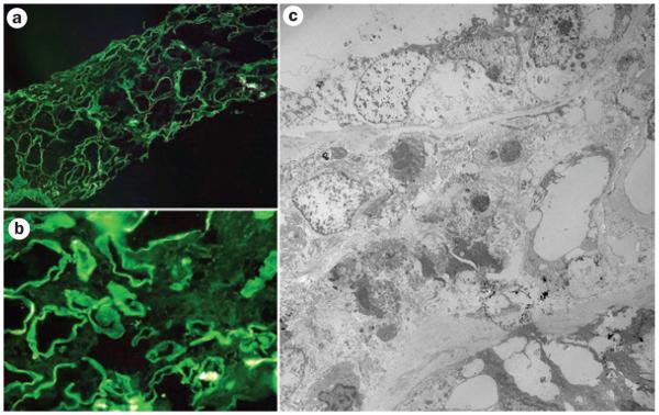
Figure 5.
Immunofluorescence and electron microscopy evaluation of tubulointerstitial damage associated with circulating monoclonal free light chains. a | and b | show examples of immunofluorescence for κ light chains. Linear staining along the tubular basement membranes represents κ light chains (negative for λ light chains). c | Electron microscopy findings of prominent tubular damage and interstitial inflammatory infiltrate. Focal punctate electron-dense material around tubular basement membranes represents deposits of free light chains.

