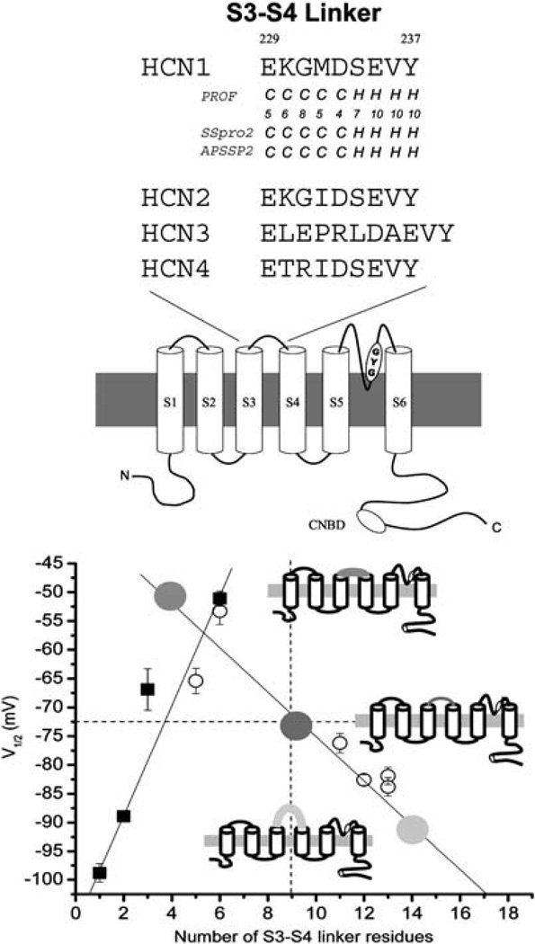
Figure 1.
(a) Schematic representation of a monomeric subunit of HCN channels. The approximate locations of the TVGYG motif and the cyclic nucleotide-binding domain are highlighted. Sequence comparison (middle and right) of the S5–S6, the P-loops and the S4 of various HCN and K+ channels. Adapted from Siu et al.8 (b) Summary of the effects of the S3–S4 linker length on HCN1 steady-state activation (V1/2), a measure of the energetic stability for opening and closing. A strong correlation was observed between the V1/2 of the channels and their linker length. Schematics representing three HCN1 channels with shorter, wild-type (WT) and longer S3–S4 linkers are given for illustration. Adapted from Tsang et al.42

