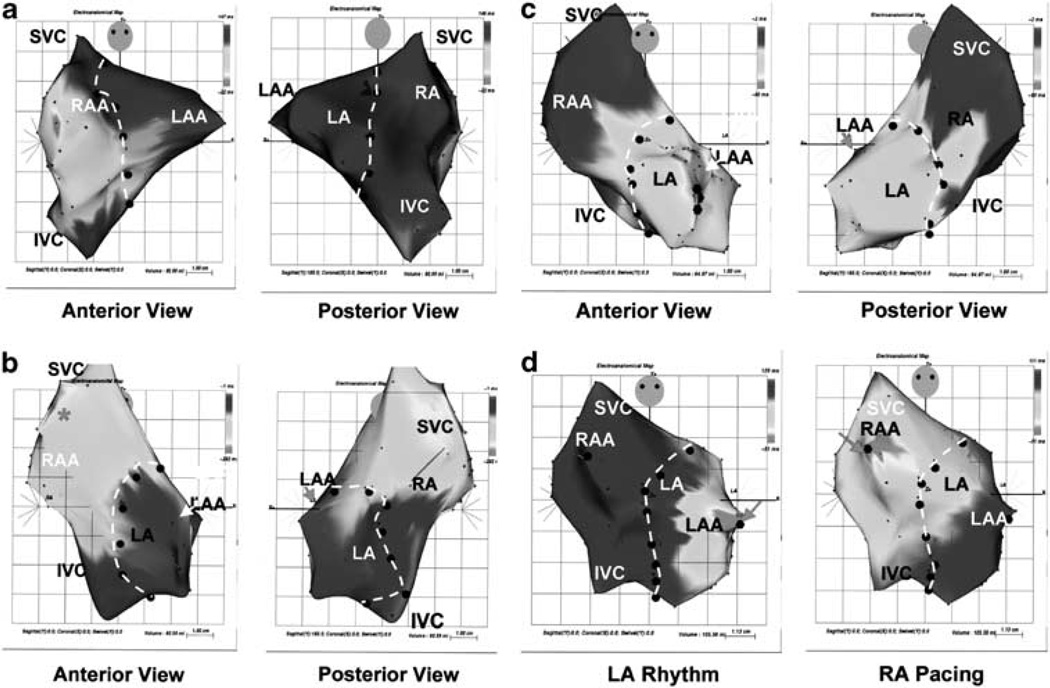
Figure 3.
Left: anterior (left) and posterior (right) views of electroanatomical mapping of (a) control and (b) a SSS swine heart that has been implanted an electronic pacemaker whose wired lead generates heart beat in the RA. (c) SSS animal after Ad-CGI-HCN1-ΔΔΔ injection demonstrate the atrial activation at the injection site in the left atrial (LA). (d) Anterior view after Ad-CGI-HCN1-ΔΔΔ injection during spontaneous LA rhythm (left) and device-supported RA pacing (right). Note that the earliest endocardial activation shifted from the left injection site to the right pacing site during device-supported RA overdrive pacing. Adapted from Tse et al.52

