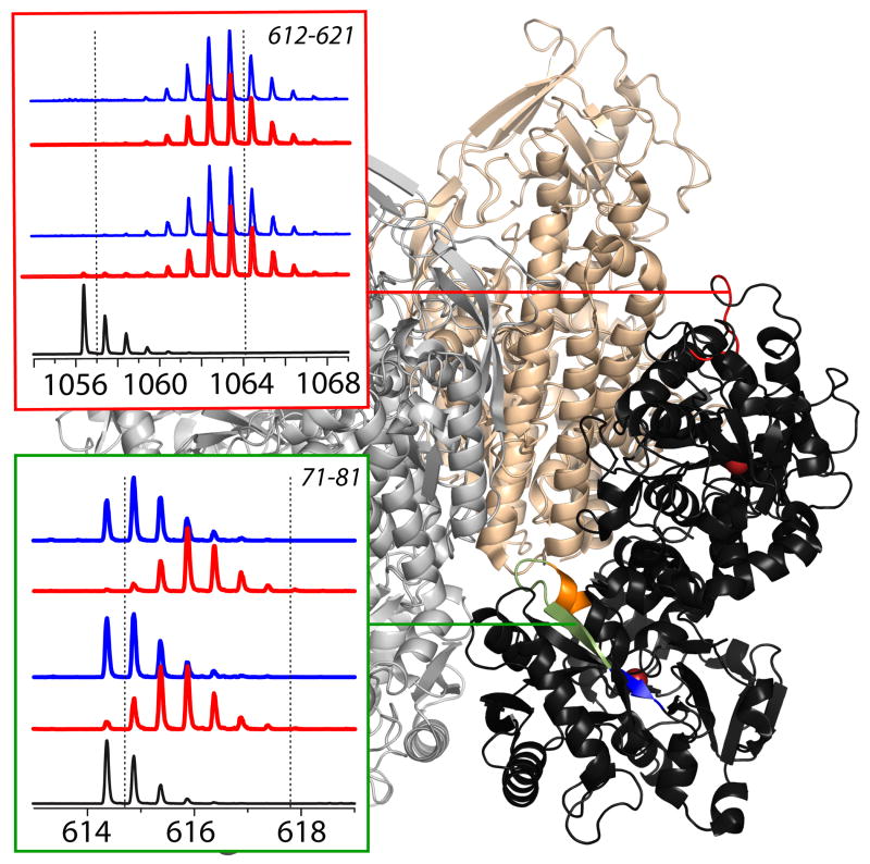
Figure 7.
Left: HDX MS within two segments of Tf in the presence (blue) and the absence (red) of the cognate receptor. The exchange was carried out by diluting the protein stock solution 1:10 in exchange solution (100 mM NH4HCO3 in D2O, pH adjusted to 7.4) and incubating for a certain period of time as indicated on each diagram followed by rapid quenching (lowering pH to 2.5 and temperature to near 0°C). The black trace at the bottom of each diagram shows unlabeled protein and the dotted lines represent the end-point of the exchange reaction. Location of these segments in Tf is shown within the context of the low-resolution structure of Tf/TfR [69].

