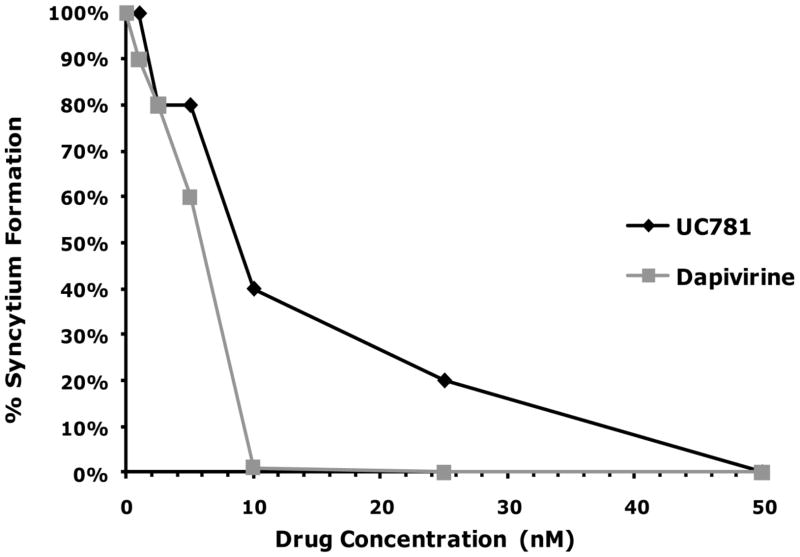
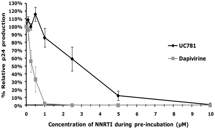
Figure 2.
(A) MT-2 cells were exposed to varying concentrations of dapivirine or UC781, and then immediately inoculated with HIV-1 (25 ng HIV-1 p24/well) in the continued presence of the drug. The extent of infection was evaluated four days after virus addition by microscopic observation of HIV-1 induced syncytium formation. Dapivirine showed greater direct antiviral potency than UC781. Dapivirine EC50 is 5 nM and UC781 EC50 is 10 nM. (B) Uninfected MT-2 cells were incubated with various concentrations of dapivirine or UC781 for 18 hours. Exogenous drug was removed by extensive washing of the cells with culture medium. The washed cells were infected with equivalent infectious doses of HIV-1 (25 ng HIV-1 p24/well). The extent of viral infection was determined four days later by microscopic observation of HIV-1 induced syncytium formation and validated by measuring the level of HIV-1 p24 antigen in the cell-free culture supernatants. Both dapivirine and UC781 were able to establish a protective barrier to infection in MT-2 cells following drug pretreatment and subsequent exposure to HIV-1 in the absence of exogenous drug. Dapivirine again showed greater potency than UC781.


