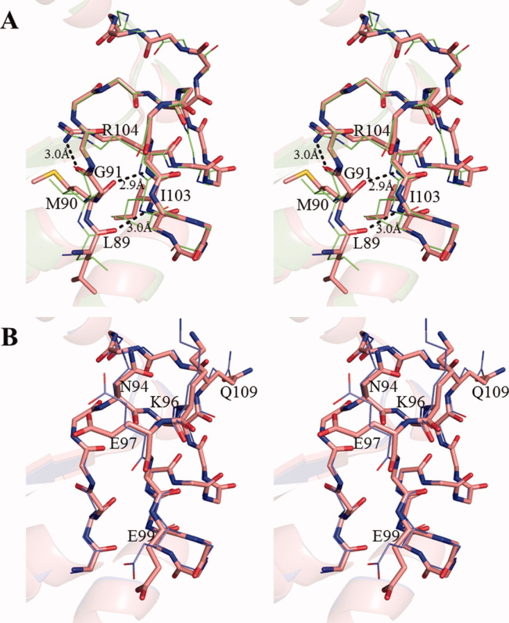
Figure 4.

Stereo view of the Kpn loop. (A), Structural comparison of the Kpn loop of Form 1 crystal (red) and EcNDK (PDB ID: 2HUR, green). These structures are superposed based on the main chain atoms. Residues (L89, M90, Q91, I103, and R104) that form the intramolecular hydrogen bonds are shown with the side chains, and the side chains of other residues are omitted. The hydrogen bonds in Form 1 crystal are shown by black dotted line. (B), Structural comparison of Kpn loop of the wild-type HaNDK (red) and the E134A mutant HaNDK (blue). The wild-type and E134A mutant HaNDKs are superposed based on the main chain atoms. N94, K96, E97, E99, and Q109 are shown with the side chains, and the side chains of other residues are omitted.
