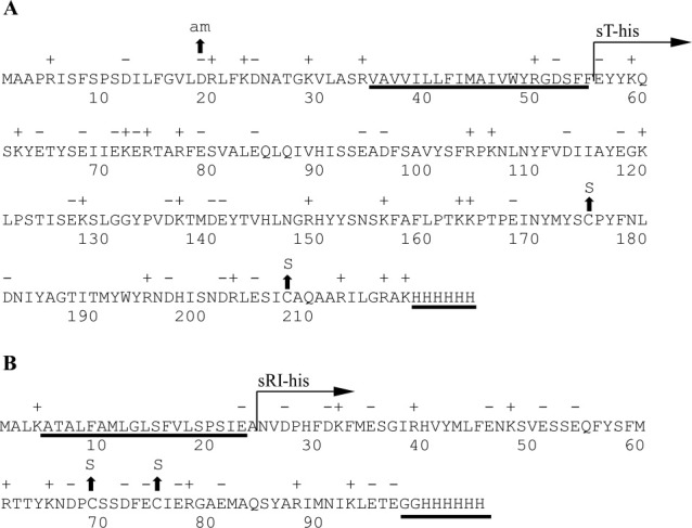
Figure 1.

The primary structure of T4 T and RI. A: T4 T. The predicted TMD and location of the C-terminal oligohistidine tag are underlined. The region of T cloned into pET11a and purified is labeled with “sT-his” (see “Materials and Methods”). Location of the amber and cysteine-to-serine substitution mutants are indicated by arrows. B: T4 RI. The SAR domain and C-terminal oligohistidine tag are underlined. The region of RI cloned into pET11a and purified is labeled with “sRI-his” (see “Materials and Methods”). Location of cysteine-to-serine substitution mutants are indicated by arrows.
