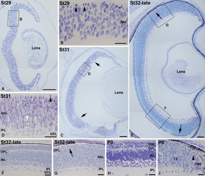
Fig. 2.

Toluidine blue-stained semi-thin transverse sections showing emergence of the retinal layers in the developing small-spotted catshark retina. Boxed areas in (A, C, E) are shown at higher magnification in (B, D, F, G). (A, B) At St29 retinal lamination was absent and the retina was occupied by a neuroepithelium with abundant mitotic figures in the scleral surface (arrowheads in B). (C, D) At St31 retinal lamination was restricted to the central retina (delimited by arrows in C), showing a GCL and an external NbL with abundant mitotic figures in the scleral-most region (black arrowhead in D) and sparse extra-ventricular mitosis (white arrowhead in D). These nuclear layers are separated by a non-cellular layer, the presumptive IPL. (E–G) At St32-late, the two plexiform layers were clearly distinguished (E–G) except in the peripheral retina, where only the presumptive GCL, IPL and external NbL were observable (E, G). (H, I) At hatching (P0) the typical cytoarchitecture of the adult retina was clearly observable (H), with the exception of the peripheral-most region, occupied by the CMZ, and a special tissue denominated transition zone, where the OPL was not evident (I). Sparse mitotic figures were observable in the peripheral retina (arrowhead in I). Scale bars: 100 μm (A, C); 200 μm (E); 25 μm (B, D, F–I). CMZ, ciliary marginal zone; GCL, ganglion cell layer; INL, inner nuclear layer; IPL, inner plexiform layer; NbL, neuroblastic layer; ONL, outer nuclear layer; OPL, outer plexiform layer; TZ, transition zone.
